Digital Posters
Fat & Metabolism I
ISMRM & SMRT Annual Meeting • 15-20 May 2021

| Concurrent 4 | 15:00 - 16:00 |
3831.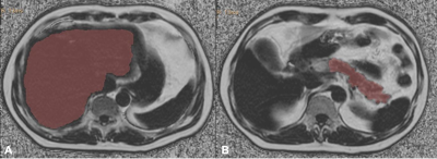 |
The relationship between hepatic and pancreatic steatosis and cardiometabolic risk factors
Qin-He Zhang1, Li-Zhi Xie2, and Ai-Lian Liu1
1The First Affiliated Hospital of Dalian Medical University, Dalian, China, 2MR Research, Beijing, China
This study assessed the correlation between hepatic and pancreatic steatosis and cardiometabolic risk factors. The results showed that hepatic and pancreatic FF were associated with specific risk factors, and the correlation coefficient varied by sex. This plays an important role in our further understanding about the association between ectopic fat deposition and cardiometabolic risk factors. Hepatic and pancreatic steatosis might have important implication for prevention cardiometabolic risk factors.
|
|||
3832.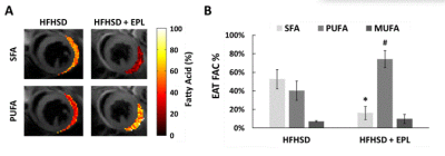 |
Cardiac MRI detects a reduced volume and anti-inflammatory fatty acid composition of epicardial adipose tissue in eplerenone-treated obese mice
Soham A Shah1, Brent French1, Matthew Wolf1, and Fred Epstein1
1University of Virginia, Charlottesville, VA, United States
Epicardial adipose tissue (EAT) volume and fatty acid composition (FAC) have significant roles in EAT-mediated coronary vascular inflammation. Using MRI of EAT volume and FAC, we tested the hypothesis that eplerenone reduces EAT volume and alters its FAC in a mouse model of obesity. We show that eplerenone significantly reduces EAT volume after 30 weeks on a high-fat high-sucrose diet compared to untreated mice. In addition, the EAT FAC is shifted from saturated fatty acid dominant in untreated mice to poly-unsaturated fatty acid dominant in eplerenone-treated mice. The results suggest that eplerenone has an anti-inflammatory role in obesity-related EAT.
|
|||
3833.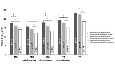 |
Deflazacort & prednisone/prednisolone treatment in Duchenne muscular dystrophy: Disease trajectory differences modeled using MR biomarkers
Ishu Arpan1, William D Rooney1, Rebecca J Willcocks2, Alison M Barnard2, Sean C Forbes2, William T Triplett2, Eric Baetscher1, Michael J Daniels2, Glenn A Walter2, and Krista Vandenborne2
1Oregon Health & Science University, Portland, OR, United States, 2University of Florida, Gainesville, FL, United States
Duchenne muscular dystrophy (DMD) is a genetic disease characterized by rapidly progressing weakness and degeneration of the skeletal muscles. This study presents a modeling approach using MR biomarkers to characterize disease progression in individuals with DMD receiving deflazacort, prednisone/prednisolone, or no corticosteroid treatment. The findings from a sample of convenience compiled from the ImagingDMD study demonstrate that 1) corticosteroid treatment slows DMD disease progression, with deflazacort slowing progression to a greater extent than prednisone/prednisolone, and 2) the slow-progressing muscles in DMD are more sensitive to treatment effects, and hence, could be better candidates to assess therapeutic response in clinical trials.
|
|||
3834.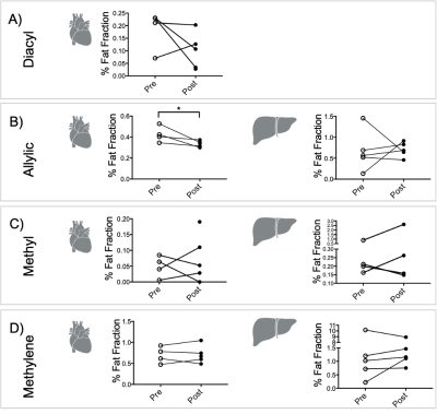 |
Cardiac and Hepatic Metabolism Modulation using L-carnitine injection: 1H Magnetic Resonance Spectroscopy Study
Dragana Savic1, Ferenc E. Mózes1, Matthew K. Burrage1, Leanne Hodson1, Stefan Neubauer1, Michael Pavlides1, Damian J. Tyler1, and Ladislav Valkovic1
1University of Oxford, Oxford, United Kingdom
This study investigated the in vivo effects on acetylcarnitine levels in the heart and the liver after an injection of L-carnitine. Carnitine acts as a buffer of acetyl-CoA units in the mitochondria, as well as facilitating transport of fatty acids. We show that an injection of L-carnitine can alter cardiac lipid levels in the in vivo healthy human. However, even though L-carnitine injection shows high cardiac variability in acetylcarnitine levels, hepatic variability of acetylcarnitine levels were reduced. Further studies are needed to confirm the dose and time-frame of a response in acetylcarnitine levels after L-carnitine injections.
|
|||
3835.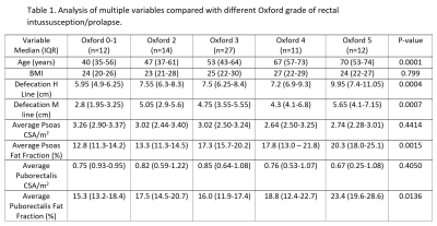 |
MRI-Based Fat Fraction of Psoas and Puborectalis Muscle Increases with Severity of Rectal Prolapse
Jonathan Philip Lam1, Leila Neshatian2, Brooke Gurland3, and Vipul Sheth4
1Radiology, Stanford University, Palo Alto, CA, United States, 2Gastroenterology, Stanford University, Palo Alto, CA, United States, 3General Surgery, Stanford University, Palo Alto, CA, United States, 4Stanford University, Palo Alto, CA, United States
Pelvic floor disorders (PFDs) are a very common group of clinical conditions that affect nearly 50% of women aged 80 years or older. While pelvic floor disorders are thought to be associated with pelvic floor muscle weakness, there has not been an established correlation between sarcopenia and PFDs. This single institution, retrospective study evaluates the effect of decreased muscle quantity/quality on severity of pelvic organ prolapse. Our findings suggest increased psoas and puborectalis muscle fat fraction are associated with higher grades of pelvic organ prolapse.
|
|||
3836.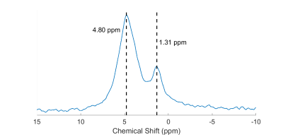 |
Natural abundance deuterium MRS of the human calf and T1 measurements with a surface coil at 3 T
Robin A. Damion1,2,3, Daniel J. Cocking3,4, Brett Haywood3,4, Matthew S. Brook2,5,6, Paul L. Greenhaff2,5,6, Philip J. Atherton1,2,5, Dorothee P. Auer1,2,3, and Richard Bowtell2,3,4
1School of Medicine, University of Nottingham, Nottingham, United Kingdom, 2NIHR Nottingham Biomedical Research Centre/Nottingham Clinical Research Facilities, University of Nottingham, Nottingham, United Kingdom, 3Sir Peter Mansfield Imaging Centre, University of Nottingham, Nottingham, United Kingdom, 4School of Physics & Astronomy, University of Nottingham, Nottingham, United Kingdom, 5MRC-Versus Arthritis Centre for Musculoskeletal Ageing Research, University of Nottingham, Nottingham, United Kingdom, 6School of Life Sciences, University of Nottingham, Nottingham, United Kingdom
Deuterium spectra of the human calf were obtained at natural abundance, at 3 T field strength. Two peaks with a chemical shift separation of 3.5 ppm were observed, corresponding to water and lipids, and their relaxation times T1 and T2* were measured using a transceive surface coil. Utilising the complex data to fit spectral lines, including independent phases for each peak, and fitting the complex inversion-recovery data enabled measurements of T1 which could be frustrated by inversion-time-dependent phases caused by RF imperfections. The results indicate that such measurements in humans are possible despite the low natural abundance of 2H.
|
|||
3837.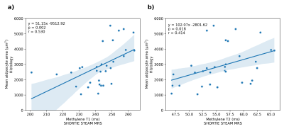 |
Characterization of the triglyceride fatty acid composition and adipocyte size in human adipose tissue using SHORTIE STEAM: in vitro validation
Stefan Ruschke1, Julius Honecker2, Dominik Weidlich1, Claudine Seeliger2, Olga Prokopchuk3, Josef Ecker4, Marcus R. Makowski1, Hans Hauner2, and Dimitrios C. Karampinos1
1Department of Diagnostic and Interventional Radiology, School of Medicine, Technical University of Munich, Munich, Germany, 2Else Kröner Fresenius Center for Nutritional Medicine, Technical University of Munich, Freising, Germany, 3Department of Surgery, Klinikum rechts der Isar, School of Medicine, Technical University of Munich, Munich, Munich, Germany, 4ZIEL Institute for Food & Health; Research Group Lipid Metabolism, Technical University of Munich, Freising, Germany
Short-TR multi-TI multi-TE (SHORTIE) STEAM is a promising technique for the characterization of adipose tissue since it allows the non-invasive simultaneous probing of triglyceride fatty acid composition and triglyceride T1- and T2-relaxation times. The present in vitro study of human adipose tissue aims at the validation of the SHORTIE-assessed triglyceride fatty acid composition with gas chromatography–mass spectrometry and investigates potential correlations of the triglyceride relaxation properties with adipocyte cell size from histology.
|
|||
3838.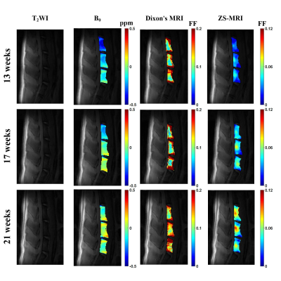 |
Early Detection of Increased Marrow Adiposity with Age Using Z-Spectral MRI
Zimeng Cai1,2, Quan Tao1,2, Alessandro Scotti3,4, Peiwei Yi1,2, Yanqiu Feng1,2, and Kejia Cai3,4
1School of Biomedical Engineering, Southern Medical University, Guangzhou, Guangdong, China, 2Guangdong Provincial Key Laboratory of Medical Image Processing, Southern Medical University, Guangzhou, Guangdong, China, 3The Department of Radiology and Bioengineering, College of Medicine, University of Illinois at Chicago, Chicago, IL, United States, 4Department of Bioengineering, University of Illinois at Chicago, Chicago, IL, United States
To investigate the capability of Z-spectral magnetic resonance imaging (ZS-MRI) for the quantification of marrow adipose tissue (MAT) content in rat’s lumbar spine and to monitor the MAT changes due to age. First, three sets of water-oil mixed phantoms prepared with varying fat fractions (FF) were scanned and the quantification results were compared to 1H-MRS and Dixon’s MRI methods. Then, ZS-MRI was used to longitudinally monitor the adiposity in the lumbar spine of healthy-young rats at 13, 17 and 21 weeks along with 1H-MRS and Dixon’s MRI. The changes over time in FF of MAT were confirmed by histology staining.
|
|||
3839.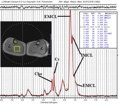 |
Healthy human aging and sex dimorphism in bone-muscle cross talk: a 1H MRS investigation in bone marrow and muscle of legs.
Silvia Capuani1,2, Roberto Coccurello3, Riccardo De Feo4, Lorenzo Rossi5, Giulia Tuttobene5, Emanuele Agrimi5, Clelia Raso5, Federico Giove2,4, and Umberto Tarantino6
1Physics Dpt Sapienza, CNR ISC, Rome, Italy, 2Santa Lucia foundation, Rome, Italy, 3CNR ISC, Rome, Italy, 4Museo Storico della Fisica e Centro Studi e Ricerche Enrico Fermi, Rome, Italy, 5Physics Dpt., Sapienza University, Rome, Italy, 6Tor Vergata University, PTV, Rome, Italy Aim of this study was to identify relevant changes in the metabolic profile of bone-marrow and muscle during healthy aging in both men and women. Towards this goal, single-voxel MRS with STEAM at TE=6ms and T2 evaluation of each resonance was performed. Bone-marrow fatty-acids quantification show a clear sex dimorphism with normal aging while no significant differences were found in the muscle metabolites of female and male subjects. On the other hand, metabolite-T2 values were different in male and female resonances and significant T2 differences among 36+, 36-52 and 52+ group, finding higher T2 in older group were observed. |
|||
3840. |
The relationship between preperitoneal fat and liver and pancreatic steatosis
Qin-He Zhang1, Li-Zhi Xie2, and Ai-Lian Liu1
1The First Affiliated Hospital of Dalian Medical University, Dalian, China, 2MR Research, Beijing, China
This study assessed the correlation between preperitoneal fat and hepatic and pancreatic steatosis. The results showed that hepatic FF and pancreatic FF were strongly associated with preperitoneal fat fraction and area. This plays an important role in our further understanding of fat distribution and ectopic fat deposition.
|
|||
3841.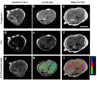 |
Noninvasive in vivo assessment of liver fat on a mouse model of diet-induced fatty liver disease using 3-Tesla MRI.
Orlando Aristizabal1, Lakshmi Arivazhagan1, AnnMarie Schmidt1, Edward A Fisher 1, Kathryn Moore1, and Youssef Zaim Wadghiri1
1NYU Grossman School of Medicine, New York, NY, United States
Nonalcoholic fatty liver disease (NAFLD) is the most common cause of liver disease in the Western The development of preclinical mouse models is essential in the development of virtual biopsy protocols to assess the essential features of NAFLD. In this report we have examined the feasibility of visualizing and mapping the fat fraction in mice under 2 high fat diets and one control diet using a preclinical 3-Tesla MRI with a RARE-Dixon sequence. We show that at 3T fat and water can be separated and percentage maps show a 23% and 39% increase in liver fat over the control mouse.
|
|||
3842.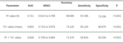 |
Evaluation of mDIXON Quant imaging in the assessment of disease activity in Thyroid-associated Ophthalmopathy(TAO)
Gang Liu1, Rongrong Zhu2, Ruoshui Ha2, and Xiuzheng Yue3
1People’s Hospital of Fuyang, Fuyang, China, 2People’s Hospital of Ningxia Hui Autonomous Region, Yinchuan, China, 3Philips Healthcare, Beijing, China
To prospectively study the changes of adipose content, adipose tissue edema and fibrosis in the orbit of Thyroid-associated ophthalmopathy (TAO) by mDixon-Quant imaging and investigate their application to TAO activity assessment. The result showed T2* values may be a promising marker for assessing the activity of TAO.
|
|||
3843.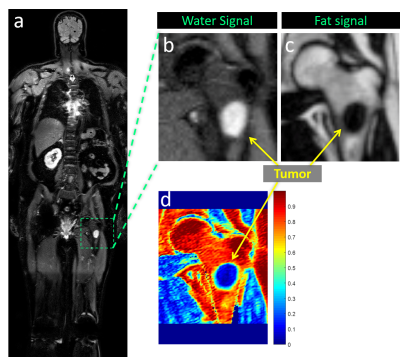 |
Fat Fraction Quantification in TSE based 2-point Dixon Acquisition: Application to Whole-body MRI
Sheng-Qing Lin1, Sebastian Fonseca1, Durga Udayakumar1, Neela Batthula1, Ivan E. Dimitrov2,3, Ivan Pedrosa1, and Ananth J. Madhuranthakam1
1Radiology, UT Southwestern Medical Center, Dallas, TX, United States, 2Advanced Imaging Research Center, UT Southwestern Medical Center, Dallas, TX, United States, 3Philips, Dallas, TX, United States
Mapping fat fraction (FF) has been proposed as a biomarker of treatment response in multiple myeloma. Whole-body imaging using 2-point Dixon methods offer efficient acquisitions but may suffer from less accurate FF quantification than multi-echo Dixon. Using our previously developed whole-body MR sequence with 2-point turbo spin-echo (TSE) Dixon, we generated accurate FF maps using with a fat masking algorithm. Results from a fat-water phantom and in vivo images show that 2-point TSE-Dixon with the fat masking algorithm performed comparably to multi-echo Dixon, facilitating accurate FF maps in whole-body MRI.
|
|||
3844.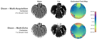 |
Three-Point Dixon Abdominal Water/Fat Separation using a Lower-Field 0.75T MRI
Christian Guenthner1, Hannes Dillinger1, Peter Boernert2,3, and Sebastian Kozerke1
1University and ETH Zurich, Zurich, Switzerland, 2Philips Research, Hamburg, Germany, 3Leiden University Medical Center, Leiden, Netherlands
Water/fat separation can be performed by exploiting chemical shift, which is proportional to field strength. Hence, on lower-field MRI systems (0.1 … 1T), the absolute resonance frequency difference is reduced compared to clinical high-field MRI, raising the question if water/fat separation can still be reliably performed despite the proximity of the resonances. In this work, we examined the feasibility of Dixon-type water/fat separation in the abdomen on a 0.75T lower-field MRI system. We show that Cartesian multi-acquisition and multi-echo as well as spiral multi-acquisition sequences provide reliable water/fat separation despite the reduced water/fat resonance shift of approximately 108 Hz.
|
|||
3845.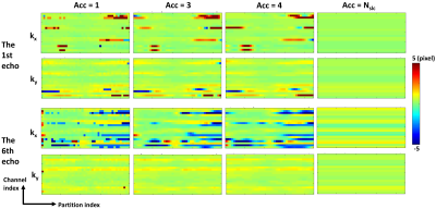 |
Improved Efficiency of Free-Breathing Stack-of-Radial MRI for Liver Fat and R2* Quantification Using Accelerated k-space Shift Calibration
Xiaodong Zhong1, Tess Armstrong2,3, Chang Gao2,3, Marcel Dominik Nickel4, Fei Han1, Brian Dale5, Holden Wu2,3, Peng Hu2,3, and Vibhas Deshpande6
1MR R&D Collaborations, Siemens Medical Solutions USA, Inc., Los Angeles, CA, United States, 2Radiological Sciences, University of California Los Angeles, Los Angeles, CA, United States, 3Physics and Biology in Medicine Interdepartmental Program, University of California Los Angeles, Los Angeles, CA, United States, 4MR Application Predevelopment, Siemens Healthcare GmbH, Erlangen, Germany, 5MR R&D Collaborations, Siemens Medical Solutions USA, Inc., Cary, NC, United States, 6MR R&D Collaborations, Siemens Medical Solutions USA, Inc., Austin, TX, United States
For liver proton density fat fraction (PDFF) and R2* quantification, free-breathing three-dimensional stack-of-radial MRI may perform a calibration scan for k-space shift correction. This study proposes an accelerated k-space shift calibration method. The proposed method was validated in a PDFF/R2* phantom with vendor-provided ground truth. Preliminary in vivo results demonstrated good agreement in the liver PDFF/R2* compared to the reference breath-hold Cartesian MRI. This proposed method enabled a 3-fold reduction in calibration time (20 seconds or more) for the in vivo protocols in this study. It may allow more efficient free-breathing PDFF/R2* mapping in patient populations with breath-hold difficulties.
|
|||
3846.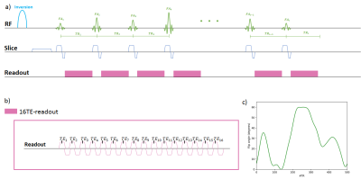 |
On the effect of fat spectrum complexity in Dixon MR Fingerprinting
Elizabeth Huaroc1, Dominik Weidlich1, Thomas Amthor2, Peter Koken2, Manuel Baumann2, Kilian Weiss3, Marcus Makowski1, Benedikt Schwaiger4, Mariya Doneva2, and Dimitrios Karampinos1
1Department of Diagnostic and Interventional Radiology, School of Medicine, Technical University of Munich, Munich, Germany, 2Philips Research Lab, Hamburg, Germany, 3Philips Healthcare, Hamburg, Germany, 4Department of Diagnostic and Interventional Neuroradiology, Technical University of Munich, Munich, Germany
Dixon Magnetic Resonance Fingerprinting (Dixon-MRF) has been emerging for achieving efficient fat suppression in body applications of MRF. Fat quantification using traditional Dixon imaging relies on a pre-calibrated proton density fat spectrum. The fat spectrum is known to be composed of different fat peaks, which have different relaxation times and are affected by J-coupling modulations. Existing Dixon-MRF methods perform water-fat separation assuming a constant pre-calibrated fat spectrum and not accounting how the complex fat spectrum varies along the MRF flip angle train. The present work proposes a framework to characterize the effect of fat spectrum complexity in Dixon-MRF.
|
|||
3847. |
Deep Learning-Based Liver Fat and R2* Mapping with Uncertainty Estimation using Self-Gated Free-Breathing Stack-of-Radial MRI
Shu-Fu Shih1,2, Sevgi Gokce Kafali1,2, Tess Armstrong1, Xiaodong Zhong3, Kara L. Calkins4, and Holden H. Wu1,2
1Radiological Sciences, University of California, Los Angeles, Los Angeles, CA, United States, 2Bioengineering, University of California, Los Angeles, Los Angeles, CA, United States, 3Siemens Medical Solutions USA, Inc., Los Angeles, CA, United States, 4Pediatrics, University of California, Los Angeles, Los Angeles, CA, United States
Self-gated free-breathing multi-echo stack-of-radial MRI quantifies liver fat and R2*. However, data undersampling due to motion self-gating can degrade the image quality and quantification accuracy. Previous methods required either longer scan time or computationally expensive constrained reconstruction. In this work, a deep learning-based two-stage network was developed to suppress undersampling artifacts and rapidly generate quantitative fat and R2* maps with a pixel-wise uncertainty map. The proposed method achieved accurate fat and R2* mapping and reduced the computational time by two orders of magnitude versus constrained reconstruction. The uncertainty map can be used to detect regions with potential quantification errors.
|
|||
 |
3848.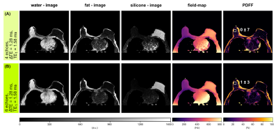 |
Optimal experimental design for quantitative water, fat and silicone separation using a variable projection method with 4 or 6 echoes at 3T
Jonathan K. Stelter1, Christof Boehm1, Stefan Ruschke1, Maximilian N. Diefenbach1,2, Mingming Wu1, Kilian Weiss3, Tabea Borde1, Stephan Metz1, Marcus R. Makowski1, and Dimitrios C. Karampinos1
1Department of Diagnostic and Interventional Radiology, School of Medicine, Technical University of Munich, Munich, Germany, 2Division of Infectious Diseases and Tropical Medicine, University Hospital, LMU Munich, Munich, Germany, 3Philips Healthcare, Hamburg, Germany
Patients with silicone breast implants undergoing MR mammography are examined with a water- and fat-suppressed sequence to verify the implant’s integrity. In parallel, chemical shift encoding-based water–fat separation has been recently used for quantifying breast water-fat composition to assess breast density and for extracting magnetic susceptibility maps to detect breast calcifications. In this work, a water–fat–silicone separation method is developed for a joint estimation of all three components and applied with NSA-optimized echo times to simulated single-voxel data, scanner phantom and in vivo data showing robust species separation and fat fraction mapping.
|
||
3849. |
Stereological Modeling of Hepatic Steatosis and Estimating Independent R2* for Water and Fat using Monte Carlo Simulations
Utsav Shrestha1, Marie Van Der Merwe2, Nirman Kumar3, Eddie Jacobs4, Sanjaya Satapathy5, Cara Morin6, and Aaryani Tipirneni-Sajja1,6
1Biomedical Engineering, The University of Memphis, Memphis, TN, United States, 2College of Health Sciences, The University of Memphis, Memphis, TN, United States, 3Computer Science, The University of Memphis, Memphis, TN, United States, 4Electrical & Computer Engineering, The University of Memphis, Memphis, TN, United States, 5North Shore University Hospital/Northwell Health, Manhasset, NY, United States, 6St. Jude Children’s Research Hospital, Memphis, TN, United States
Multi-spectral fat-water models assume either single or independent R2* for water (R2*W) and fat (R2*F) for assessment of steatosis. However, any incorrect assumptions in the signal model will produce errors in R2* and fat fraction (FF) calculations. In this study, we developed a Monte Carlo–based approach for generating steatosis models by characterizing fat morphology from histology and synthesizing MRI signal to estimate independent R2*W and R2*F and compare with in-vivo studies. Our results show that R2*W and R2*F are slightly different at low FFs and both demonstrate a positive correlation with FF with slopes of R2*W-FF similar to in-vivo calibrations.
|
|||
3850.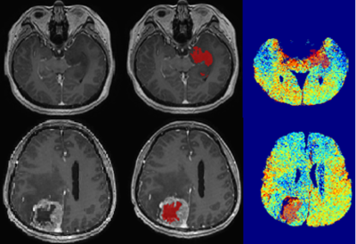 |
MRI lipid signatures and SREBPs gene expression profiling for characterisation of glioma: A preliminary study
Pohchoo Seow1, K Rashid2, Vairavan Narayanan 3, Azlina Ahmad-Annuar 4, Jeannie Wong2, Li Kuo Tan2, K Ibrahim4, and Norlisah Ramli2
1Diagnostic Radiology, Singapore General Hospital, Singapore, Singapore, 2Department of Biomedical Imaging, University of Malaya, Kuala Lumpur, Malaysia, 3Department of Surgery, University of Malaya, Kuala Lumpur, Malaysia, 4Department of Biomedical Science, University of Malaya, Kuala Lumpur, Malaysia
We explored the relationships between MRI lipid-imaging signatures and sterol regulatory element binding proteins (SREBPs) gene expression profiling of ten histologically proven glioma patients to elucidate the underlying inter- & intra-tumoural lipid distribution changes. The high expression of SREBPs was observed for grade II and III implied reprogramming of lipid metabolism. The identification of imaging biomarkers that can reflect changes in the genetic profiles of SREBP offers a new avenue for personalised medicine and the development of new therapeutic targets.
|
The International Society for Magnetic Resonance in Medicine is accredited by the Accreditation Council for Continuing Medical Education to provide continuing medical education for physicians.