Weekend Course
Antenatal & Pediatric MRI Challenges
ISMRM & SMRT Annual Meeting • 15-20 May 2021

| Concurrent 1 | 14:30 - 15:15 | Moderators: Antenatal & Pediatric Renal & Abdominal: Claudia Hillenbrand & Minhui Ouyang Antenatal & Childhood Cardiac & Spinal: Julio Garcia Flores & Elka Miller |
| Antenatal & Pediatric Renal & Abdominal | ||
 |
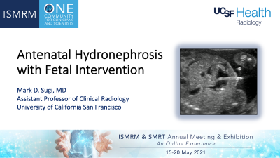
Antenatal Hydronephrosis with Fetal Intervention
Mark Sugi, Mark Sugi
Antenatal urinary tract dilation (UTD) occurs in 1-2% of fetuses and may be caused by multiple genitourinary anomalies, although mild forms are often transient. UTD can be detected as early as the first trimester as megacystis. Differential diagnosis for fetal UTD can be refined via assessment of the kidneys, ureters, bladder, sex, and amniotic fluid volume. Early detection allows for potential interventions aimed at increasing amniotic fluid volume to allow for critical pulmonary development. While UTD is often detected by ultrasound, fetal MRI has a role in further characterizing renal function and delineating the anatomy of complex urinary tract anomalies.
|
|
 |
Neonatal/Pediatric MR Urography with Renal Perfusion
Cara Morin
MRU provides a thorough anatomic and functional assessment of the urinary tract in children, allowing detailed evaluation of the renal parenchyma, collecting systems and ureters, and the bladder, while also providing both static and dynamic functional information. As such, MRU has the potential to be contributory to the evaluation of a wide variety of pediatric urologic abnormalities. With new motion-robust dynamic post-contrast sequences, quantitative assessment of renal perfusion is becoming increasingly accessible as a clinical technique.
|
|
| Antenatal/Neonatal Cystic & Solid Masses
Teresa Victoria
In this session we will review common and uncommon feral lesions of the chest, with postnatal follow up. Diagnosis pearls will be discussed with an emphasis on MR imaging. Also discussed will be cystic and solid lesions of the fetal abdomen, with postnatal follow up. |
||
 |
Pediatric Neuroblastoma: Diagnosis & Staging
Alexander Towbin
Neuroblastoma is the most common extracranial soft tissue malignancy in children. In 2004, the International Neuroblastoma Risk Group (INRG) published a new staging system designed to standardize the presurgical staging of neuroblastoma. The INRG staging system developed a series of 20 different image-defined risk factors (IDRF) that confer additional surgical risk and upstage a patient from L1 to L2 disease. Radiologists should be familiar with the different IDRFs and the standard definitions used assess patients with neuroblastoma.
|
|
| Antenatal & Childhood Cardiac & Spinal | ||
| Antenatal MR Techniques: Congenital Heart Disease
David Lloyd
|
||
| Neonatal/Pediatric Cardiac 4D Flow & Fractional Flow Reserve
Joshua Robinson
|
||
| Antenatal Spinal Pathology with Fetal Surgical Intervention Video Permission Withheld
Usha Nagaraj
This talk will review the pathology of open spinal dysraphism and Chiari II malformation, the current surgical techniques for repair and the imaging criteria for fetal surgery. Relevant imaging findings on fetal and postnatal MRI in this population will be reviewed.
|
||
| Neonatal/Pediatric Spinal Pathology
Helen Branson
|
||
The International Society for Magnetic Resonance in Medicine is accredited by the Accreditation Council for Continuing Medical Education to provide continuing medical education for physicians.